Arthritis / Acute Gout Attack
Gout is a form of arthritis, hence it causes pain and discomfort in the joints. A typical gout attack is characterized by the sudden onset of severe pain, swelling, warmth, and redness of a joint. The clinical presentation of acute gouty arthritis is not subtle with very few mimics other than a bacterial infection.
The joint most commonly involved in gout is the first metatarsophalangeal joint (the big toe), and is called podagra. Any joint may be involved in a gout attack (and it may be more than one) with the most frequent sites being in the feet, ankles, knees, and elbows.
An acute gout attack will generally reach its peak 12-24 hours after onset, and then will slowly begin to resolve even without treatment. Full recovery from a gout attack (without treatment) takes approximately 7-14 days.
An accurate and colorful discription of a gout attack was elegantly written in 1683 by Dr. Thomas Sydenham who was himself a sufferer of gout:
The victim goes to bed and sleeps in good health. About 2 o’clock in the morning, he is awakened by a severe pain in the great toe; more rarely in the heel, ankle or instep. This pain is like that of a dislocation, and yet the parts feel as if cold water were poured over them. Then follows chills and shiver and a little fever. The pain which at first moderate becomes more intense. With its intensity the chills and shivers increase. After a time this comes to a full height, accommodating itself to the bones and ligaments of the tarsus and metatarsus. Now it is a violent stretching and tearing of the ligaments– now it is a gnawing pain and now a pressure and tightening. So exquisite and lively meanwhile is the feeling of the part affected, that it cannot bear the weight of bedclothes nor the jar of a person walking in the room.
Chronic Tophaceous Gout
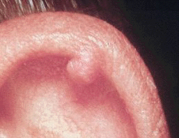
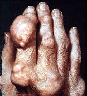
Some patients only experience acute gout attacks which may be limited to 1-2 times per year (or even 1-2 times in lifetime). However, for some patients, gout can be a chronic, relapsing problem with multiple severe attacks that occur at short intervals and without complete resolution of inflammation between attacks. This form of gout, called chronic gout, can cause significant joint destruction and deformity and may be confused with other forms of chronic inflammatory arthritis such as rheumatoid arthritis. Frequently, uric acid tophi (hard, uric acid deposits under the skin) are present and contribute to bone and cartilage destruction. Tophi are diagnostic for chronic tophaceous gout. Tophi can be found around joints, in the olecranon bursa, or at the pinna of the ear. With treatment, tophi can be dissolved and will completely disappear over time.


Asymptomatic Hyperuricemia
It is important to recognize that although almost uniformly all patients with gout have hyperuricemia (high levels of uric acid in the blood)…all patients with hyperuricemia do not have gout. Although most patients will have elevated levels of uric acid in the blood for many years before having their first gout attack, there is no current recommendation for treatment during this period in the absence of clinical signs or symptoms of gout. This is termed ‘asymptomatic hyperuricemia’. The risk of a gout attack increases with increasing uric acid levels, but many patients will have attacks with “normal” levels of uric acid and some will never have an attack despite very high levels of uric acid.
Diagnosis of Gout
A diagnosis of gout can be made with the documentation of the presence of uric acid crystals in synovial fluid or from a tophaceous deposit. In the setting of an acute gout attack, aspiration of joint fluid (by using a needle to draw fluid out of the swollen joint) and examination of the fluid under polarized light can yield the definitive diagnostic finding of needle shaped negatively-birefringent uric acid crystals (yellow when parallel to the axis of polarization). Intracellular crystals within a neutrophil are characteristic during an acute attack.
As the clincal features of acute gout and a septic joint (bacterial infection) can be very similar, arthrocentesis is important to rule out infection by sending the joint fluid for culture in these circumstances. Importantly, gout and infection can co-exist in the same joint (they are not mutually exclusive) and consideration should be made for sending joint fluid for culture even in a patient with an established history of gout if they are at risk for infection.
Tophi can be aspirated or the tophaceous material expressed and examined under polarize microscopy as well to confirm a diagnosis of chronic tophaceous gout.
Serum uric acid concentrations may be supportive of a diagnosis of gout, but alone the presence of hyperuricemia or normal uric acid concentrations do not confirm or rule out the diagnosis of gout as frequently uric acid levels may be normal during an acute gout attack.

